File:Plasmacytoma1.jpg

Summary
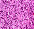
<a href="https://en.wikipedia.org/wiki/Micrograph" class="extiw" title="w:Micrograph">Micrograph</a> of a <a href="https://en.wikipedia.org/wiki/plasmacytoma" class="extiw" title="w:plasmacytoma">plasmacytoma</a>. <a href="https://en.wikipedia.org/wiki/H%26E_stain" class="extiw" title="w:H&E stain">H&E stain</a>.
The micrograph shows abundant (malignant) <a href="https://en.wikipedia.org/wiki/plasma_cell" class="extiw" title="w:plasma cell">plasma cells</a> with the occasional Mott cell, a plasma cell with an intracytoplasmic <a href="https://en.wikipedia.org/wiki/Russell_body" class="extiw" title="w:Russell body">Russell bodies</a> (an eosinophilic uniformly staining membrane bound body which contains immunoglobulin).
Other features of plasmacytomas (not apparent on the image) are:
- a prominent perinuclear hof (large <a href="https://en.wikipedia.org/wiki/Golgi_body" class="extiw" title="w:Golgi body">Golgi bodies</a>), and
- Dutcher bodies (intranuclear inclusions).
The plasma cells have the characteristic "clockface nuclei".
<a href="https://en.wikipedia.org/wiki/multiple_myeloma" class="extiw" title="w:multiple myeloma">Multiple myeloma</a> (which is diagnosed using several clinical criteria) is, histologically, a plasmacytoma.
Related images
-
<a href="//commons.wikimedia.org/wiki/File:Plasmacytoma1.jpg" class="image"><img alt="" src="https://upload.wikimedia.org/wikipedia/commons/thumb/4/4e/Plasmacytoma1.jpg/120px-Plasmacytoma1.jpg" width="120" height="102" srcset="https://upload.wikimedia.org/wikipedia/commons/thumb/4/4e/Plasmacytoma1.jpg/180px-Plasmacytoma1.jpg 1.5x, https://upload.wikimedia.org/wikipedia/commons/thumb/4/4e/Plasmacytoma1.jpg/240px-Plasmacytoma1.jpg 2x" data-file-width="3179" data-file-height="2690"></a>
Uncropped.
-
<a href="//commons.wikimedia.org/wiki/File:Plasmacytoma_ultramini1.jpg" class="image"><img alt="" src="https://upload.wikimedia.org/wikipedia/commons/thumb/1/10/Plasmacytoma_ultramini1.jpg/120px-Plasmacytoma_ultramini1.jpg" width="120" height="88" srcset="https://upload.wikimedia.org/wikipedia/commons/thumb/1/10/Plasmacytoma_ultramini1.jpg/180px-Plasmacytoma_ultramini1.jpg 1.5x, https://upload.wikimedia.org/wikipedia/commons/thumb/1/10/Plasmacytoma_ultramini1.jpg/240px-Plasmacytoma_ultramini1.jpg 2x" data-file-width="950" data-file-height="700"></a>
Cropped.
Licensing
Lua error in package.lua at line 80: module 'strict' not found.
File history
Click on a date/time to view the file as it appeared at that time.
| Date/Time | Thumbnail | Dimensions | User | Comment | |
|---|---|---|---|---|---|
| current | 08:06, 8 January 2017 | | 3,179 × 2,690 (3.29 MB) | 127.0.0.1 (talk) | <a href="https://en.wikipedia.org/wiki/Micrograph" class="extiw" title="w:Micrograph">Micrograph</a> of a <b><a href="https://en.wikipedia.org/wiki/plasmacytoma" class="extiw" title="w:plasmacytoma">plasmacytoma</a></b>. <a href="https://en.wikipedia.org/wiki/H%26E_stain" class="extiw" title="w:H&E stain">H&E stain</a>. <p>The micrograph shows abundant (malignant) <a href="https://en.wikipedia.org/wiki/plasma_cell" class="extiw" title="w:plasma cell">plasma cells</a> with the occasional <i>Mott cell</i>, a plasma cell with an intracytoplasmic <a href="https://en.wikipedia.org/wiki/Russell_body" class="extiw" title="w:Russell body">Russell bodies</a> (an eosinophilic uniformly staining membrane bound body which contains immunoglobulin). </p> <p>Other features of plasmacytomas (not apparent on the image) are: </p> <ul> <li>a prominent perinuclear hof (large <a href="https://en.wikipedia.org/wiki/Golgi_body" class="extiw" title="w:Golgi body">Golgi bodies</a>), and</li> <li>Dutcher bodies (intranuclear inclusions).</li> </ul> <p>The plasma cells have the characteristic "clockface nuclei". </p> <p><a href="https://en.wikipedia.org/wiki/multiple_myeloma" class="extiw" title="w:multiple myeloma">Multiple myeloma</a> (which is diagnosed using several clinical criteria) is, histologically, a plasmacytoma. </p> <h2><span class="mw-headline" id="Related_images">Related images</span></h2> <ul class="gallery mw-gallery-traditional"> <li class="gallerybox" style="width: 155px"><div style="width: 155px"> <div class="thumb" style="width: 150px;"><div style="margin:24px auto;"><a href="//commons.wikimedia.org/wiki/File:Plasmacytoma1.jpg" class="image"><img alt="" src="https://upload.wikimedia.org/wikipedia/commons/thumb/4/4e/Plasmacytoma1.jpg/120px-Plasmacytoma1.jpg" width="120" height="102" srcset="https://upload.wikimedia.org/wikipedia/commons/thumb/4/4e/Plasmacytoma1.jpg/180px-Plasmacytoma1.jpg 1.5x, https://upload.wikimedia.org/wikipedia/commons/thumb/4/4e/Plasmacytoma1.jpg/240px-Plasmacytoma1.jpg 2x" data-file-width="3179" data-file-height="2690"></a></div></div> <div class="gallerytext"> <p>Uncropped. </p> </div> </div></li> <li class="gallerybox" style="width: 155px"><div style="width: 155px"> <div class="thumb" style="width: 150px;"><div style="margin:31px auto;"><a href="//commons.wikimedia.org/wiki/File:Plasmacytoma_ultramini1.jpg" class="image"><img alt="" src="https://upload.wikimedia.org/wikipedia/commons/thumb/1/10/Plasmacytoma_ultramini1.jpg/120px-Plasmacytoma_ultramini1.jpg" width="120" height="88" srcset="https://upload.wikimedia.org/wikipedia/commons/thumb/1/10/Plasmacytoma_ultramini1.jpg/180px-Plasmacytoma_ultramini1.jpg 1.5x, https://upload.wikimedia.org/wikipedia/commons/thumb/1/10/Plasmacytoma_ultramini1.jpg/240px-Plasmacytoma_ultramini1.jpg 2x" data-file-width="950" data-file-height="700"></a></div></div> <div class="gallerytext"> <p>Cropped. </p> </div> </div></li> </ul> |
- You cannot overwrite this file.
File usage
The following 2 pages link to this file: